TriTeQ3: Compact trite Q 3D camera system for binocular microscopes
Project summary
The NIHR HealthTech Research Centre in Devices, Digital and Robotics (NIHR HRC-DDR) collaborated with Vision Engineering Ltd, a UK-based ergonomic and digital microscope company to evaluate a 3D camera system developed for use on healthcare binocular microscopes.
Clinical need
Binocular optical microscopes are widely used in healthcare settings, providing the user with a 3D view of the area of interest. They have changed little in design and function, and the user is often seated in an uncomfortable position for long periods. Images from the microscope can be projected onto display screens to allow other members of the healthcare team see the procedure for training, remote referrals and can also be recorded. However, the projected image is often in 2D, and existing 3D displays require the viewer to wear additional glasses which reduce the intensity of the viewed image.
The solution
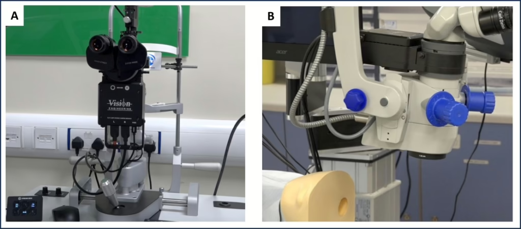
Vision Engineering (VE) have developed a novel, stereoscopic camera system for industrial inspections that can project a 3D image onto a display screen without the need for additional glasses and with no loss of signal. VE have now produced a compact version, (Compact TriTeQ3, CTTQ), for use in healthcare settings, fitting the stereo camera into the optical path of slit-lamp or surgical binocular microscopes (Figure 1).
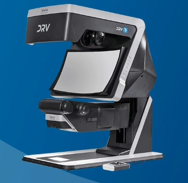
Images captured using the stereoscopic camera can be displayed on an ergonomic, stereoscopic display unit. A large format Deep Reality viewer (DRV) display, and a compact (CTTQ) version are available (Figure 2).
How we supported
The NIHR HRC-DDR collaborated on applications for grant funding for product development, provided regulatory and commercialisation support, and delivered human factors validation (formative usability) studies of the technology.
NIHR HRC-DDR supported by:
- Identifying and partnering on grant funding applications to secure a 12 month Innovate UK grant.
- Planning, setting up and moderating two formative usability studies to assess the ease of use, training requirements and instructions for use of prototype versions of CTTQ in a realistic, simulated environment.
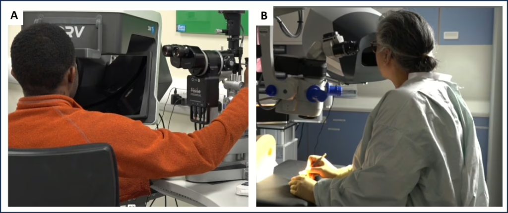
- Recruited 13 ophthalmologists and optometrists to a formative usability study of the CTTQ fitted to a slit lamp microscope. This included liaising with Vision Sciences at Aston University to set up and evaluate the system in their clinic rooms.
- Recruited 7 ophthalmic surgeons to a formative usability study of the CTTQ system fitted to a binocular surgical microscope in the MD-TEC operating theatre to perform a simulated cataract removal surgery.
- Filming of the testing sessions using static, high- definition cameras and conducting post-study questionnaires and interviews.
- Compiling comprehensive reports (to IEC 62366 standard) and edited video for both usability studies to summarise the outcomes and participants feedback.
- Worked with Vision Engineering and Device Access UK to discuss the requirements for and undertake a health economic analysis of the technology.
Outcomes
Feedback from the formative usability sessions was positive with participants reporting the system to be easy and straightforward to use. A number of potential improvements were suggested and summarised for the developers including optimisation of the 3D screen position, simplifying the video recording process and reducing the impact the camera had on reaching the controls of the slit lamp.
The work with Device Access provided an expert/insider insight into the economic climate for introduction of novel 3D camera system to healthcare market.
The usability studies and health economic analysis have provided useful information to guide further development of the system. A project plan is in preparation to identify and apply for continuation funding, and to identify clinical partners to achieve a clinical study.
Latest case studies

Addressing the Unmet Need in DiabeticNeuropathy: Insights…
Dynamis MedTech is advancing research into painful diabetic peripheral neuropathy (pDPN) with the Sensetic device,…
TriTeQ3: Compact trite Q 3D camera system…
Project summary The NIHR HealthTech Research Centre in Devices, Digital and Robotics (NIHR HRC-DDR) collaborated…
SurePulse Medical Ltd – Development of a…
Project summary The NIHR HealthTech Research Centre in Devices, Digital and Robotics (NIHR HRC-DDR, previously…